
January 2024
January’s Ultrasound of the Month goes to Dr. Bui!
He captured a very subtle acute cholecystitis. The clip included demonstrates cholelithiasis with trace pericholecystic fluid (why I feel it is a subtle scan) with a normal CBD. The GB wall also happened to be normal. I am hopeful that one day our surgeons will trust our ED ultrasounds to expedite patient flow, instead of waiting for a comprehensive ultrasound.
Most struggle with finding the CBD. Tips to find this:
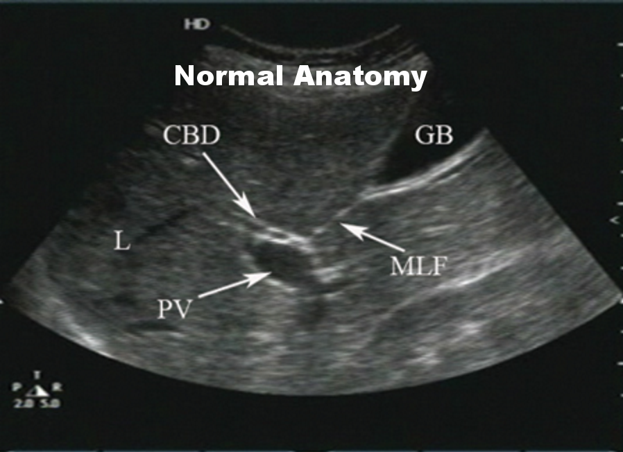
1) In long axis view, follow the gallbladder down to 3 structures. There is a line here known as the median lobar fissure (MLF). This line connects the gallbladder to the portal triad.
A look at normal GB anatomy on US.
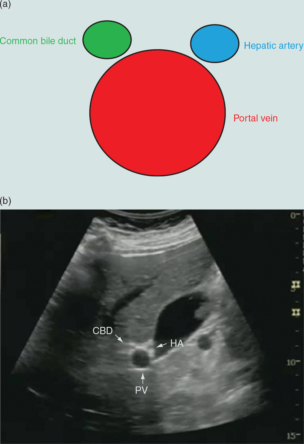
2) Once you find the triad, place color over the structures. Have the patient hold their breath.
(The portal triad looks like a Mickey Mouse face in short axis)
3) In color mode, the structure that does not light up with color is the CBD.
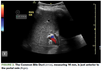